- Компании
- Takeda. О компании, буклеты, каталоги, контакты
- Olympus. О компании, буклеты, каталоги, контакты
- Boston Scientific. О компании, буклеты, каталоги, контакты
- Pentax. О компании, буклеты, каталоги, контакты
- Fujifilm & R-Farm. О компании, буклеты, каталоги, контакты
- Erbe. О компании, буклеты, каталоги, контакты
- Еще каталоги
- Мероприятия
- Информация
- Обучение
- Классификации
- Атлас
- Quiz
- Разделы
- Пациенту
QR-код этой страницы
Для продолжения изучения на мобильном устройстве ПРОСКАНИРУЙТЕ QR-код с помощью спец. программы или фотокамеры мобильного устройства
Статьи: Азбука Эндоскописта. Эндоскопические классификации для описания и оценки патологических изменений толстой кишки при колоноскопии
Полный текст статьи:
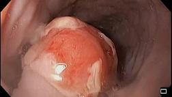
- Макроскопическая классификация поверхностных эпителиальных образований (тип роста образования) – Парижская классификация
The Paris endoscopic classification of superficial neoplastic lesions: esophagus, stomach, and colon/ Participants in the Paris Workshop, Paris, France, 2002//Gastrointestinal endoscopy. – 2003. – V 58(6 Suppl). – P. S 3–43. Update on the paris classification of superficial neoplastic lesions in the digestive tract. Review/ Endoscopic Classification Review Group// Endoscopy. – 2005. – V 37(6). – P. 570–8.
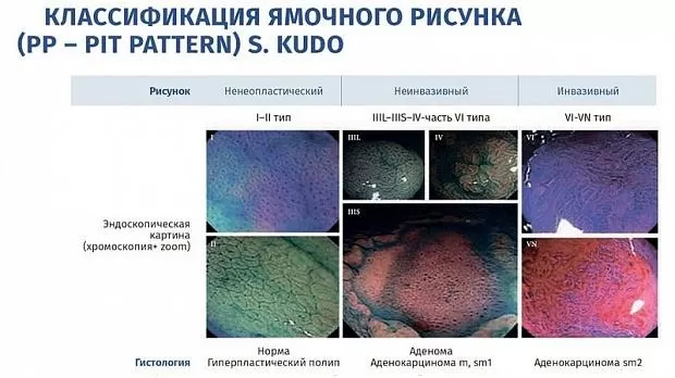
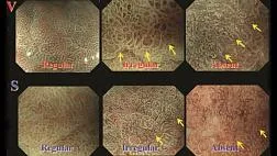
- Описание ямочного рисунка поверхности – по классификации S. Kudo3, а при подозрении на наличие зубчатого образования – дополнительное описание ямочного рисунка II–O типа (по T. Kimura)
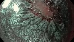
- Описание капиллярного (сосудистого) рисунка поверхности –по классификации Y. Sano
Ikematsu H., Matsuda T., Emura F. et al. Efficacy of capillary pattern type IIIA/IIIB by magnifying narrow band imaging for estimating depth of invasion of early colorectal neoplasms. BMC Gastroenterol, 2010, vol. 10, pp. 33.
Iwatate M., Ikumoto T., Hattori S. et al. NBI and NBI combined with magnifying colonoscopy. Diagn Ther Endosc, 2012, 173269.
- NICE и JNET ЭНДОСКОПИЧЕСКАЯ КЛАССИФИКАЦИЯ JNET YASUSHI SANO, DAIZEN HIRATA, YUTAKA SAITO (ОБРАЗОВАТЕЛЬНОЕ ВИДЕО)
Ijspeert JEG, Bastiaansen BaJ, van Leerdam ME, et al. Development and validation of the WASP classification system for optical diagnosis of adenomas, hyperplastic polyps and sessile serrated adenomas/polyps. Gut 2016;65:963-70.
The BASIC classification for colorectal polyp characterization uses Blue Light Imaging System and stands for BLI Adenoma Serrated International Classification, i.e. a classification of colon adenomas, including serrated lesions based on BLI technology, which can be read in detail in the March issue of Endoscopy (Bisschops et al., Endoscopy. 2018 Mar; 50(3):211–20).

- классификации в эндоскопии
- Азбука Эндоскописта. Эндоскопические классификации для описания и оценки патологических изменений толстой кишки при колоноскопии
- Классификация WASP: дифференциальная диагностика гиперпластических полипов, зубчатых аденом и обычных аденом толстой кишки
- Классификация и диагностика гастритов и гастропатий
- Номенклатурная классификация медицинских изделий 3.15 Эндоскопы гастроэнтерологические
- Z-линия пищевода. Классификация ZAP
- Парижская классификация
- Классификации ЭУС критериев хронического панкреатита
- ЭНДОСКОПИЧЕСКАЯ КЛАССИФИКАЦИЯ JNET YASUSHI SANO, DAIZEN HIRATA, YUTAKA SAITO (ОБРАЗОВАТЕЛЬНОЕ ВИДЕО)

Рекомендуемые статьи
При эндоскопическом исследовании в случае бронхоэктазов в стадии ремиссии выявляется
частично диффузный бронхит I степени воспаления
Активируйте PUSH уведомления в браузер
Отключите PUSH уведомления в браузер
Содержание
Интернет магазин
Популярное
- О нас
- Правовые вопросы
- Политика
обработки персональных
данных EndoExpert.ru - Связаться с нами
- Стать партнером
© 2016-2022 EndoExpert.ru

Вы находитесь в разделе предназначенном только для специалистов (раздел для пациентов по ссылке). Пожалуйста, внимательно прочитайте полные условия использования и подтвердите, что Вы являетесь медицинским или фармацевтическим работником или студентом медицинского образовательного учреждения и подтверждаете своё понимание и согласие с тем, что применение рецептурных препаратов, обращение за той или иной медицинской услугой, равно как и ее выполнение, использование медицинских изделий, выбор метода профилактики, диагностики, лечения, медицинской реабилитации, равно как и их применение, возможны только после предварительной консультации со специалистом. Мы используем файлы cookie, чтобы предложить Вам лучший опыт взаимодействия. Файлы cookie позволяют адаптировать веб-сайты к вашим интересам и предпочтениям.
Я прочитал и настоящим принимаю вышеизложенное, хочу продолжить ознакомление с размещенной на данном сайте информацией для специалистов.






.jpg)

.png)



Комментарии