Просмотренные публикации

QR-код этой страницы

Для продолжения изучения на мобильном устройстве ПРОСКАНИРУЙТЕ QR-код с помощью спец. программы или фотокамеры мобильного устройства

Случайный выбор

данная функция, случайным образом выбирает информацию для Вашего изучения,
запустите выбор нажав кнопку ниже

Обратная связь
Напишите нам

Поделитесь своими идеями по улучшению нашей работы.
Прикрепить файл или скриншот удалить
Закрытая часть портала предназначена для только для работников здравоохранения. Оставив свой e-mail и специалищацию, Вы подтверждаете, что являетесь работником здравоохранения и что Вы ознакомились с текстом и поняли его.


Сообщение об ошибке
Что улучшить?

Поделитесь своими идеями по улучшению нашей работы.
Прикрепить файл или скриншот удалить
Закрытая часть портала предназначена для только для работников здравоохранения. Оставив свой e-mail и специалищацию, Вы подтверждаете, что являетесь работником здравоохранения и что Вы ознакомились с текстом и поняли его.


Главная ► Статьи ► Эндоскопическая шкала EREFS при оценке эозинофильного эзофагита (Hirano, 2012 г.)

Статьи: Эндоскопическая шкала EREFS при оценке эозинофильного эзофагита (Hirano, 2012 г.)

Анонс:

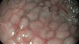
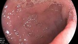
Эозинофильный эзофагит (ЭоЭ) — это хроническое иммуноопосредованное заболевание пищевода, характеризующееся симптомами эзофагеальной дисфункции и выраженной эозинофильной инфильтрацией слизистой оболочки органа.

Диагностика ЭоЭ основывается на

  • · данных клинической картины заболевания (дисфагия, эпизоды вклинения пищи в пищевод, боль в грудной клетке без связи с глотанием), а также
  • · на совокупности эндоскопических и гистологических признаков.

Критерием установления диагноза служит эозинофильная инфильтрация слизистой оболочки пищевода с плотностью эозинофилов ≥ 15 в поле зрения микроскопа при большом увеличении (×400) по крайней мере в одном из биоптатов (около 60 эозинофилов в мм2).

Вспомогательное значение для диагноза имеют:

  • · исследование общего IgE,
  • · эозинофилия периферической крови,
  • · результаты кожных аллергопроб.

1 / 168
Оцените материал:

Полный текст статьи:

Коротко

EREFS (Eosinophilic Esophagitis Endoscopic Reference Score) — стандартизированная система описания и градации основных эндоскопических признаков эозинофильного эзофагита (ЭоЭ).

Аббревиатура EREFS расшифровывается как Edema — Rings — Exudates — Furrows — Stricture: отек, кольца, экссудаты, продольные борозды и стриктура. Система используется для стандартизированной эндоскопической оценки активности ЭоЭ и наблюдения изменений эндоскопической картины в динамике.

Таблица системы

Компонент EREFS Критерий Балл
E — Edema
Отек
Отсутствует: сосудистый рисунок отчетливо виден 0
Присутствует: потеря четкости или отсутствие сосудистого рисунка 1
R — Rings
Кольца
Отсутствуют 0
Легкие: едва заметные циркулярные кольца 1
Умеренные: отчетливые кольца, не препятствующие проведению стандартного диагностического эндоскопа 2
Выраженные: отчетливые кольца, не позволяющие провести стандартный диагностический эндоскоп 3
E — Exudates
Экссудаты
Отсутствуют 0
Легкие: поражено менее 10% поверхности пищевода 1
Выраженные: поражено 10% или более поверхности пищевода 2
F — Furrows
Продольные борозды
Отсутствуют 0
Присутствуют 1
S — Stricture
Стриктура
Отсутствует 0
Присутствует 1

При суммировании пяти компонентов итоговая оценка EREFS находится в диапазоне 0–8 баллов.

Как указывать в протоколе и заключении

Компоненты EREFS следует фиксировать в порядке, соответствующем аббревиатуре: Edema → Rings → Exudates → Furrows → Stricture.

Пример компактной записи:

EREFS: E1R2Ex1F1S0.

Во избежание неоднозначности две буквы E можно дополнительно расшифровать в описательной части протокола: Edema 1; Rings 2; Exudates 1; Furrows 1; Stricture 0.

Как интерпретировать результат

EREFS стандартизирует описание пяти характерных эндоскопических признаков ЭоЭ. Отек, экссудаты и продольные борозды относятся к воспалительным эндоскопическим признакам; кольца и стриктуры отражают фибростенотические изменения.

Итоговый балл формируется суммированием оценок отдельных компонентов. При динамическом наблюдении целесообразно сохранять не только общий балл, но и значения каждого компонента EREFS, поскольку одинаковая сумма может складываться из разных эндоскопических признаков.

Что это за система

Eosinophilic Esophagitis Endoscopic Reference Score (EREFS) — система стандартизированного описания и градации эндоскопических признаков эозинофильного эзофагита.

В аббревиатуру включены пять признаков:

  • E — Edema — отек;
  • R — Rings — кольца;
  • E — Exudates — экссудаты;
  • F — Furrows — продольные борозды;
  • S — Stricture — стриктура.

EREFS предназначена для унификации эндоскопической терминологии и оценки выраженности эндоскопических проявлений ЭоЭ. Эндоскопическая оценка не заменяет другие компоненты диагностики заболевания.

Ограничения и частые ошибки

Ошибка Что учитывать
Перестановка двух компонентов E Первая E в EREFS означает Edema, вторая E — Exudates.
Использование трех степеней отека В упрощенной клинической градации edema оценивается как отсутствие (0) или наличие (1).
Использование трех степеней продольных борозд В упрощенной клинической градации furrows оцениваются как отсутствие (0) или наличие (1).
Ориентация только на итоговую сумму Следует сохранять значения отдельных компонентов EREFS, поскольку они описывают разные эндоскопические признаки.
Использование EREFS как самостоятельного диагностического критерия EREFS стандартизирует эндоскопическую оценку и не является самостоятельным подтверждением диагноза ЭоЭ.

Версия классификации и модификации

Классификация была разработана и валидирована Hirano и соавт. В исследовании первоначально оценивались четыре основных эндоскопических признака — rings, furrows, exudates и edema — и дополнительные признаки, включая stricture, narrow-caliber esophagus, feline esophagus и crepe-paper esophagus.

При оценке межнаблюдательной согласованности градации edema и furrows были укрупнены, а признаки feline esophagus и narrow-caliber esophagus исключены из модифицированной системы из-за недостаточной воспроизводимости. В современной клинической записи EREFS используется последовательность Edema — Rings — Exudates — Furrows — Stricture.

Источники

  1. Hirano I., Moy N., Heckman M.G., Thomas C.S., Gonsalves N., Achem S.R. Endoscopic assessment of the oesophageal features of eosinophilic oesophagitis: validation of a novel classification and grading system. Gut. 2013;62(4):489–495. DOI: 10.1136/gutjnl-2011-301817.
  2. Dellon E.S. et al. Accuracy of the Eosinophilic Esophagitis Endoscopic Reference Score in Diagnosis and Determining Response to Treatment. Clinical Gastroenterology and Hepatology. 2016.
  3. Endoscopic approach to eosinophilic esophagitis: American Society for Gastrointestinal Endoscopy Consensus Conference. Gastrointestinal Endoscopy. 2022.

По теме


Открыт общий доступ к полному тексту статьи свернуть

Список литературы:

Литература / References 1. Трухманов А.С., Ивашкина Н.Ю. Эозинофильный эзофагит. В кн. Ивашкин В.Т. (ред.) Рациональная фармакотерапия заболеваний органов пищеварения. 2-е изд. М.: Литерра, 2011. С. 267–9 [Trukhmanov A.S., Ivashkina N.Yu. Eosinophilic esophagitis. In: Ivashkin V.T. (ed.) Rational pharmacotherapy of digestive system diseases. 2nd ed. Moscow: Literra, 2011. P. 267–9 (In Rus.)]. 2. Ивашкин В.Т., Баранская Е.К., Кайбышева В.О., Иванова Е.В. и др. Эозинофильный эзофагит: обзор литературы и описание собственного наблюдения. Рос журн гастроэнт гепатол колопроктол. 2012;22(1):71–81 [Ivashkin V.T., Baranskaya E.K., Kaibysheva V.O., Ivanova E.V. et al. Eosinophilic esophagitis: A literature review and the description of the authors’ experience. Russian Journal of Gastroenterology, Hepatology, Coloproctology. 2012;22(1):71–81 (In Rus.)]. 3. Lucendo A.J., Molina-Infante J., Arias Á. et al. Guidelines on eosinophilic esophagitis: evidence-based statements and recommendations for diagnosis and management in children and adults. United European Gastroenterol J. 2017;5(3):335–58. DOI: 10.1177/2050640616689525 4. Dellon E.S., Gonsalves N., Hirano I. et al. ACG clinical guideline: evidenced based approach to the diagnosis and management of esophageal eosinophilia and eosinophilic esophagitis. Am J Gastroenterol. 2013;108:679–92. 5. Cianferoni A., Spergel J.M., Muir A. Recent advances in the pathological understanding of eosinophilic esophagitis. Expert Rev Gastroenterol Hepatol. 2015;9(12):1501–10. DOI: 10.1586/17474124.2015.1094372 6. Hill D.A., Spergel J.M. The Immunologic Mechanisms of Eosinophilic Esophagitis. Curr Allergy Asthma Rep. 2016;16(2):9. DOI: 10.1007/s11882-015-0592-3 7. Spergel J.M. New genetic links in eosinophilic esophagitis. Genome Med. 2010;2(9):60. 8. Dellon E.S., Hirano I. Epidemiology and Natural History of Eosinophilic Esophagitis. Gastroenterology. 2018;154(2):319–32.e3. DOI: 10.1053/j.gastro.2017.06.067 9. Параскевова А.В., Шелковникова И.И., Лапина Т.Л., Трухманов А.С. и др. Пациент 68 лет и пациентка 52 лет с эозинофильной инфильтрацией пищевода. Рос журн гастроэнт гепатол колопроктол 2017;27(4):62–74 [Paraskevova A.V., Shelkovnikova I.I., Lapina T.L., Trukhmanov A.S. et al. Two cases of eosinophilic infiltration of the esophagus: 68 year-old male and 52 year-old female patients. Ross z gastroenterol gepatol koloproktol. 2017;27(4):62–74 (In Rus.)]. DOI: 10.22416/1382-4376- 2017-27-4-62-74 10. Dellon E.S. Epidemiology of eosinophilic esophagitis. Gastroenterol Clin North Am. 2014;43:201–18. 11. Prasad G.A., Alexander J.A., Schleck C.D. et al. Epidemiology of eosinophilic esophagitis over three decades in Olmsted County, Minnesota. Clin Gastroenterol Hepatol. 2009;7:1055–61. 12. Kapel R.C., Miller J.K., Torres C. et al. Eosinophilic esophagitis: a prevalent disease in the United States that affects all age groups. Gastroenterology. 2008;134:1316–21. 13. Castro Jimenez A., Gomez Torrijos E., Garcia Rodriguez R. et al. Demographic, clinical and allergological characteristics of Eosinophilic Esophagitis in a Spanish central region. Allergol Immunopathol (Madr). 2014;42:407–14. 14. van Rhijn B.D., Verheij J., Smout A.J. et al. Rapidly increasing incidence of eosinophilic esophagitis in a large cohort. Neurogastroenterol Motil. 2013;25:47–52. 15. Arias A., Perez-Martinez I., Tenias J.M. et al. Systematic review with meta-analysis: the incidence and prevalence of eosinophilic oesophagitis in children and adults in population-based studies. Aliment Pharmacol Ther. 2016;43:3–15. 16. González-Cervera J., Arias Á., Redondo-González O. et al. Association between atopic manifestations and eosinophilic esophagitis: a systematic review and meta-analysis. Ann Allergy Asth Immunol. 2017;118(5):582–90.e2. DOI: 10.1016/j.anai.2017.02.006 17. Remedios M., Campbell C., Jones D.M. et al. Eosinophilic esophagitis in adults: clinical, endoscopic, histologic findings, and response to treatment with fluticasone propionate. Gastrointest Endosc. 2006;63:3–12. 18. Croese J., Fairley S.K., Masson J.W. et al. Clinical and endoscopic features of eosinophilic esophagitis in adults. Gastrointest Endosc. 2003;58:516–22. 19. Spergel J.M., Brown-Whitehorn T.F., Beausoleil J.L. et al. 14 years of eosinophilic esophagitis: clinical features and prognosis. J Pediatr Gastroenterol Nutr. 2009;48:30–6. 20. Dellon E.S., Speck O., Woodward K. et al. Distribution and variability of esophageal eosinophilia in patients undergoing upper endoscopy. Mod Pathol. 2015;28:383–90. 21. Fiocca R., Mastracci L., Engström C. et al. Long-term outcome of microscopic esophagitis in chronic GERD patients treated with esomeprazole or laparoscopic antireflux surgery in the LOTUS trial. Am J Gastroenterol. 2010;105:1015–23. 22. Dellon E.S., Liacouras C.A., Molina-Infante J., Furuta G.T. et al. Updated International Consensus Diagnostic Criteria for Eosinophilic Esophagitis: Proceedings of the AGREE Conference. Gastroenterology. 2018;155(4):1022– 33.e10. DOI: 10.1053/j.gastro.2018.07.009 23. Lucendo A.J., Arias A., Tenias J.M. Relation between eosinophilic esophagitis and oral immunotherapy for food allergy: a systematic review with meta-analysis. Ann Allergy Asthma Immunol. 2014;113(6):624–9. DOI: 10.1016/j.anai.2014.08.004 24. Schlag C., Miehlke S., Heiseke A., Brockow K. et al. Peripheral blood eosinophils and other non-invasive biomarkers can monitor treatment response in eosinophilic oesophagitis. Aliment Pharmacol Ther. 2015 Nov;42(9):1122–30. DOI: 10.1111/apt.13386 25. Hines B.T., Rank M.A., Wright B.L., Marks L.A. et al. Minimally invasive biomarker studies in eosinophilic esophagitis: A systematic review. Ann Allergy Asthma Immunol. 2018;121(2):218–28. DOI: 10.1016/j.anai.2018.05.005 26. Pelz B.J., Wechsler J.B, Amsden K., Johnson К. et al. IgE-associated food allergy alters the presentation of pediatric eosinophilic esophagitis. Clin Exp Allergy. 2016;46(11):1431–40. 27. Hirano I., Moy N., Heckman M.G. et al. Endoscopic assessment of the oesophageal features of eosinophilic oesophagitis: validation of a novel classification and grading system. Gut. 2013;62:489–95. 28. Kim H.P., Vance R.B., Shaheen N.J. et al. The prevalence and diagnostic utility of endoscopic features of eosinophilic esophagitis: a meta-analysis. Clin Gastroenterol Hepatol. 2012;10:988–96. 29. Hirano I. Role of advanced diagnostics for eosinophilic esophagitis. Dig Dis. 2014;32(1–2):78–83. 30. Liacouras C.A., Spergel J.M., Ruchelli E. et al. Eosinophilic esophagitis: A 10-year experience in 381 children. Clin Gastroenterol Hepatol. 2005;3:1198–206. 31. Saffari H., Peterson K.A., Fang J.C. et al. Patchy eosinophil distributions in an esophagectomy specimen from a patient with eosinophilic esophagitis: Implications for endoscopic biopsy. J Allergy Clin Immunol. 2012;130:798– 800. 32. Nielsen J.A., Lager D.J., Lewin M. et al. The optimal number of biopsy fragments to establish a morphologic diagnosis of eosinophilic esophagitis. Am J Gastroenterol. 2014;109:515–20. 33. Salek J., Clayton F., Vinson L. et al. Endoscopic appearance and location dictate diagnostic yield of biopsies in eosinophilic oesophagitis. Aliment Pharmacol Ther. 2015;41:1288–95. 34. Dellon E.S., Speck O., Woodward K. et al. Distribution and variability of esophageal eosinophilia in patients undergoing upper endoscopy. Mod Pathol. 2015;28:383–90. 35. Kim H.P., Dellon E.S. An Evolving Approach to the Diagnosis of Eosinophilic Esophagitis. Gastroenterol Hepatol (N Y). 2018 Jun;14(6):358–66. 36. Saffari H., Hoffman L.H., Peterson K.A. et al. Electron microscopy elucidates eosinophil degranulation patterns in patients with eosinophilic esophagitis. J Allergy Clin Immunol. 2014;133:1728–34. 37. Capocelli K.E., Fernando S.D., Menard-Katcher C. et al. Ultrastructural features of eosinophilic oesophagitis: impact of treatment on desmosomes. J Clin Pathol. 2015;68:51–6. 38. Collins M.H. Histopathologic features of eosinophilic esophagitis and eosinophilic gastrointestinal diseases. Gastroenterol Clin North Am. 2014;43:257–68. 39. Anyane-Yeboa A., Wang W., Kavitt R.T. The Role of Allergy Testing in Eosinophilic Esophagitis. Gastroenterol Hepatol (N Y). 2018 Aug;14(8):463–9. 40. Syed A., Maradey-Romero C., Fass R. The relationship between eosinophilic esophagitis and esophageal cancer. Dis Esophagus. 2017;30(7):1–5. DOI: 10.1093/dote/ dox050 41. Lucendo A.J., Arias A., Molina-Infante J. Efficacy of proton pump inhibitor drugs for inducing clinical and histologic remission in patients with symptomatic esophageal eosinophilia: a systematic review and meta-analysis. Clin Gastroenterol Hepatol. 2016;14:13–22. 42. Molina-Infante J., Ferrando-Lamana L., Ripoll C. et al. Esophageal eosinophilic infiltration responds to proton pump inhibition in most adults. Clin Gastroenterol Hepatol. 2011;9:110–7. 43. Liacouras C.A., Furuta G.T., Hirano I. et al. Eosinophilic esophagitis: updated consensus recommendations for children and adults. J Allergy Clin Immunol. 2011;128:3– 20. 44. Gómez-Torrijos E., García-Rodríguez R., CastroJiménez A. et al. The efficacy of step-down therapy in adult patients with proton pump inhibitor-responsiveoesophageal eosinophilia. Aliment Pharmacol Ther. 2016 Feb;43(4):534–40. DOI: 10.1111/apt.13496 45. Schaefer E.T., Fitzgerald J.F., Molleston J.P. et al. Comparison of oral prednisone and topical fluticasone in the treatment of eosinophilic esophagitis: a randomized trial in children. Clin Gastroenterol Hepatol. 2008;6:165–73. 46. Tan N. Di, Xiao Y.L., Chen M.H. Steroids therapy for eosinophilic esophagitis: Systematic review and meta-analysis. J Dig Dis. 2015;16:431–42. 47. Sawas T., Dhalla S., Sayyar M. et al. Systematic review with meta-analysis: pharmacological interventions for eosinophilic oesophagitis. Aliment Pharmacol Ther. 2015;41:797–806. 48. Murali A.R., Gupta A., Attar B.M. et al. Topical steroids in eosinophilic esophagitis: systematic review and metaanalysis of placebo controlled randomized clinical trials. J Gastroenterol Hepatol. 2015;31:1111–9. 49. Chuang M.-Y.A., Chinnaratha M.A., Hancock D.G. et al. Topical steroid therapy for the treatment of eosinophilic esophagitis (EoE): a systematic review and metaanalysis. Clin Transl Gastroenterol. 2015;6:e82. 50. Straumann A., Conus S., Degen L. et al. Long-term budesonide maintenance treatment is partially effective for patients with eosinophilic esophagitis. Clin Gastroenterol Hepatol. 2011;9:400–9. 51. Greuter T., Safroneeva E., Bussmann C. et al. Maintenance Treatment Of Eosinophilic Esophagitis With Swallowed Topical Steroids Alters Disease Course Over A 5-Year Follow-Up Period In Adult Patients. Clin Gastroenterol Hepatol. 2018:S1542-3565(18)30598-6. DOI: 10.1016/j.cgh.2018.05.045 52. Eluri S., Runge T.M., Hansen J. et al. Diminishing Effectiveness of Long-Term Maintenance Topical Steroid Therapy in PPI Non-Responsive Eosinophilic Esophagitis. Clin Transl Gastroenterol. 2017;8(6):e97. DOI: 10.1038/ctg.2017.27 53. Moawad F.J., Cheatham J.G., DeZee K.J. Meta-analysis: the safety and efficacy of dilation in eosinophilic oesophagitis. Aliment Pharmacol Ther. 2013;38:713–20. 54. Kagalwalla A.F., Sentongo T.A., Ritz S. et al. Effect of six-food elimination diet on clinical and histologic outcomes in eosinophilic esophagitis. Clin Gastroenterol Hepatol. 2006;4:1097–102. 55. Arias A., Gonzalez-Cervera J., Tenias J.M. et al. Efficacy of dietary interventions for inducing histologic remission in patients with eosinophilic esophagitis: a systematic review and meta-analysis. Gastroenterology. 2014;146:1639–48. 56. Molina-Infante J., Arias A., Barrio J. et al. Four-food group elimination diet for adult eosinophilic esophagitis: A prospective multicenter study. J Allergy Clin Immunol. 2014;134:1093–99. 57. Molina-Infante J., Modolell I., Alcedo J. et al. Step-up empiric elimination diet for pediatric and adult eosinophilic esophagitis: the 2–4–6 study. United Eur Gastroenterol J. 2016;(Suppl 5):A126. 58. Gonsalves N., Yang G.-Y., Doerfler B. et al. Elimination diet effectively treats eosinophilic esophagitis in adults; food reintroduction identifies causative factors. Gastroenterology. 2012;142:1451–5. 59. Lucendo A.J., Arias A., Gonzalez-Cervera J. et al. Empiric 6-food elimination diet induced and maintained prolonged remission in patients with adult eosinophilic esophagitis: a prospective study on the food cause of the disease. J Allergy Clin Immunol. 2013;131:797–804. 60. Kagalwalla A.F., Shah A., Li B.U.K. et al. Identification of specific foods responsible for inflammation in children with eosinophilic esophagitis successfully treated with empiric elimination diet. J Pediatr Gastroenterol Nutr. 2011;53:145–9. 61. Safroneeva E., Straumann A., Coslovsky M. et al. Symptoms have modest accuracy in detecting endoscopic and histologic remission in adults with eosinophilic esophagitis. Gastroenterology. 2016;150:581–90.
10.05.20 ©
Оцените материал: Рейтинг: 8

Комментарии

Написать

При эндоскопическом исследовании в случае бронхоэктазов в стадии ремиссии выявляется

частично диффузный бронхит I степени воспаления

Интернет магазин

Работаем и учимся при поддержке

Партнеры