- Компании
- Takeda. О компании, буклеты, каталоги, контакты
- Olympus. О компании, буклеты, каталоги, контакты
- Boston Scientific. О компании, буклеты, каталоги, контакты
- Pentax. О компании, буклеты, каталоги, контакты
- Fujifilm & R-Farm. О компании, буклеты, каталоги, контакты
- Erbe. О компании, буклеты, каталоги, контакты
- Еще каталоги
- Мероприятия
- Информация
- Обучение
- Классификации
- Атлас
- Quiz
- Разделы
- Пациенту
QR-код этой страницы
Для продолжения изучения на мобильном устройстве ПРОСКАНИРУЙТЕ QR-код с помощью спец. программы или фотокамеры мобильного устройства
Статьи: Классификации применяемые при пероральной холангиоскопии для диагностики поражений желчных протоков. Роблес-Медранда 2018г.
| Авторы: | Карлос Роблес-Медранда 2018г. |
Полный текст статьи:
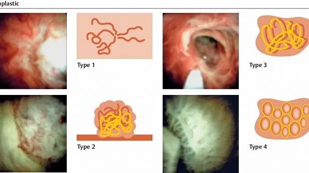
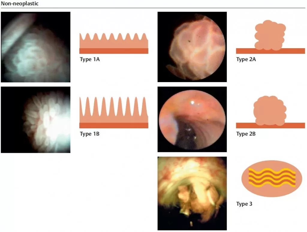
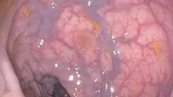
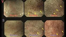
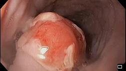
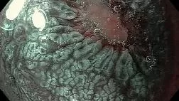
В 2018 авторами предложена классификация изменений при эндоскопической холангиоскопии, которая позволяет различить доброкачественное от злокачественного новообразования по его виду. Т.е. по макроскопической оценке протоковых образований, сосудистого рисунка, признаков воспаления и других признаков.
Авторы заключают, что новая классификационная система показывает лучшие результаты и может оказать помощь специалистам в различении доброкачественных от неопластических образований желчных путей.
|
▶ Table 1 Peroral cholangioscopy macroscopic classification system of non-neoplastic and neoplastic common bile duct lesions. | ||
|
Non-neoplastic lesions | ||
|
Type 1 |
Villous pattern |
A Micronodular, or B Villous pattern without vascularity |
|
Type 2 |
Polypoid pattern |
A Adenoma, or B Granuloma pattern without vascularity |
|
Type 3 |
Inflammatory pattern |
Regular or irregular fibrous and congestive pattern with regular vascularity |
|
Neoplastic lesions | ||
|
Type 1 |
Flat pattern |
Flat and smooth or irregular surface with irregular or spider vascularity and without ulceration |
|
Type 2 |
Polypoid pattern |
Polypoid with fibrosis and irregular or spider vascularity |
|
Type 3 |
Ulcerated pattern |
Irregular ulcerated and infiltrative pattern with or without fibrosis and with irregular or spider vascularity |
|
Type 4 |
Honeycomb pattern |
Fibrous honeycomb pattern with or without irregular or spider vascularity |

Статьи по теме
Рекомендуемые статьи
Синдром Бергмана
расстройство сердечной деятельности с изменениями ЭКГ при рефлюкс-эзофагите и аксиальной грыже
При эндоскопическом исследовании в случае бронхоэктазов в стадии ремиссии выявляется
частично диффузный бронхит I степени воспаления
Активируйте PUSH уведомления в браузер
Отключите PUSH уведомления в браузер
Содержание
Интернет магазин
Популярное
- О нас
- Правовые вопросы
- Политика
обработки персональных
данных EndoExpert.ru - Связаться с нами
- Стать партнером
© 2016-2022 EndoExpert.ru

Вы находитесь в разделе предназначенном только для специалистов (раздел для пациентов по ссылке). Пожалуйста, внимательно прочитайте полные условия использования и подтвердите, что Вы являетесь медицинским или фармацевтическим работником или студентом медицинского образовательного учреждения и подтверждаете своё понимание и согласие с тем, что применение рецептурных препаратов, обращение за той или иной медицинской услугой, равно как и ее выполнение, использование медицинских изделий, выбор метода профилактики, диагностики, лечения, медицинской реабилитации, равно как и их применение, возможны только после предварительной консультации со специалистом. Мы используем файлы cookie, чтобы предложить Вам лучший опыт взаимодействия. Файлы cookie позволяют адаптировать веб-сайты к вашим интересам и предпочтениям.
Я прочитал и настоящим принимаю вышеизложенное, хочу продолжить ознакомление с размещенной на данном сайте информацией для специалистов.







.jpg)

.png)








Комментарии