- Компании
- Takeda. О компании, буклеты, каталоги, контакты
- Olympus. О компании, буклеты, каталоги, контакты
- Boston Scientific. О компании, буклеты, каталоги, контакты
- Pentax. О компании, буклеты, каталоги, контакты
- Fujifilm & R-Farm. О компании, буклеты, каталоги, контакты
- Erbe. О компании, буклеты, каталоги, контакты
- Еще каталоги
- Мероприятия
- Информация
- Обучение
- Классификации
- Атлас
- Quiz
- Разделы
- Пациенту
QR-код этой страницы
Для продолжения изучения на мобильном устройстве ПРОСКАНИРУЙТЕ QR-код с помощью спец. программы или фотокамеры мобильного устройства
Статьи: Классификация WASP: дифференциальная диагностика гиперпластических полипов, зубчатых аденом и обычных аденом толстой кишки
Анонс:
Полный текст статьи:
Классификация WASP – эндоскопическая диагностика полипов меньше 10 мм.
До определенного времени считалось, что единственными предшественниками рака толстой кишки являются аденоматозные полипы. В связи с этим в основу разработанных эндоскопических классификаций (Kudo, NICE, JNET) был заложен принцип дифференциальной диагностики неопластических (аденоматозных) образований от не неопластических (гиперпластических) [1-3].
Однако недавно была выделена новая группа образований, являющихся предшественниками рака толстой кишки, это зубчатые аденомы (альтернативный путь канцерогенеза). Считается, что рак толстой кишки развивается из зубчатых аденом в 15-30% случаев [4, 5].
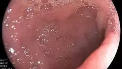
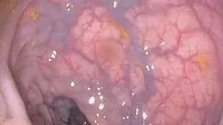
Но ввиду своего макроскопического сходства с гиперпластическими полипами зубчатые аденомы часто не распознаются врачами-эндоскопистами как неопластические образования. Также ни в одной из перечисленных классификаций (Kudo, NICE, JNET) не описан подход к их визуальной диагностике [3, 6]. Это диктует необходимость в разработке стратегии по эндоскопической диагностике данных образований.
Именно с этой целью была разработана классификация WASP [7].
Классификация WASP позволяет дифференцировать:
- 1. гиперпластический полип,
- 2. зубчатую аденому,
- 3. обычную аденому.
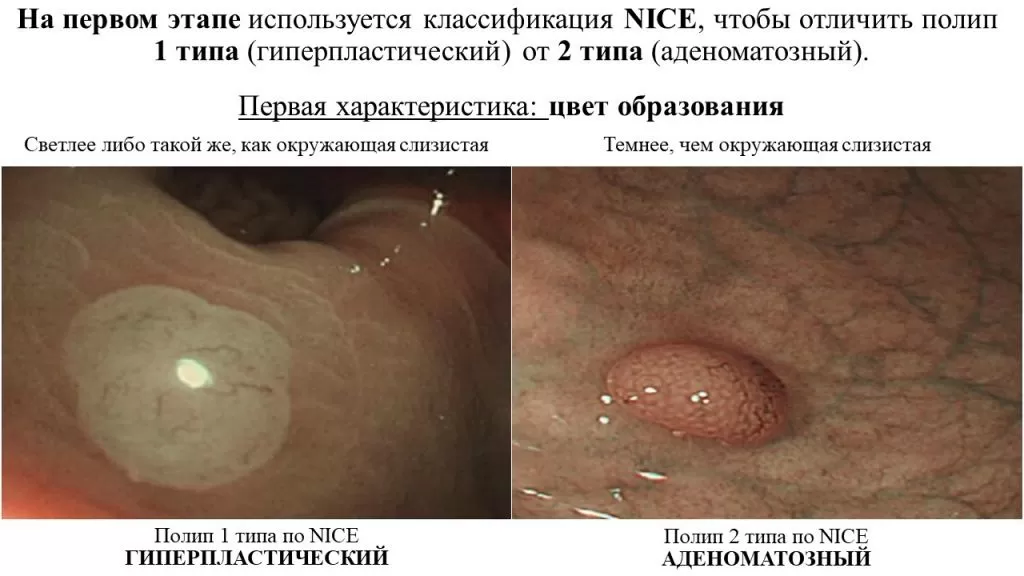
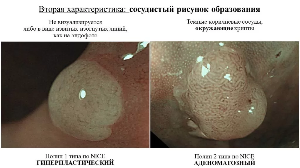
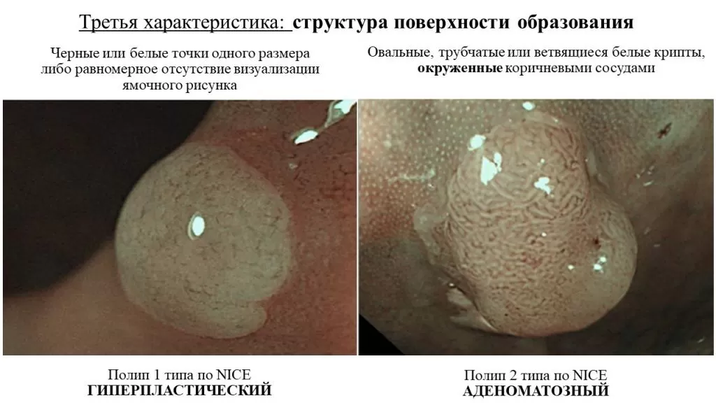
На первом этапе используется классификация NICE, чтобы отличить полип 1 типа (гиперпластический) от 2 типа (аденоматозный).
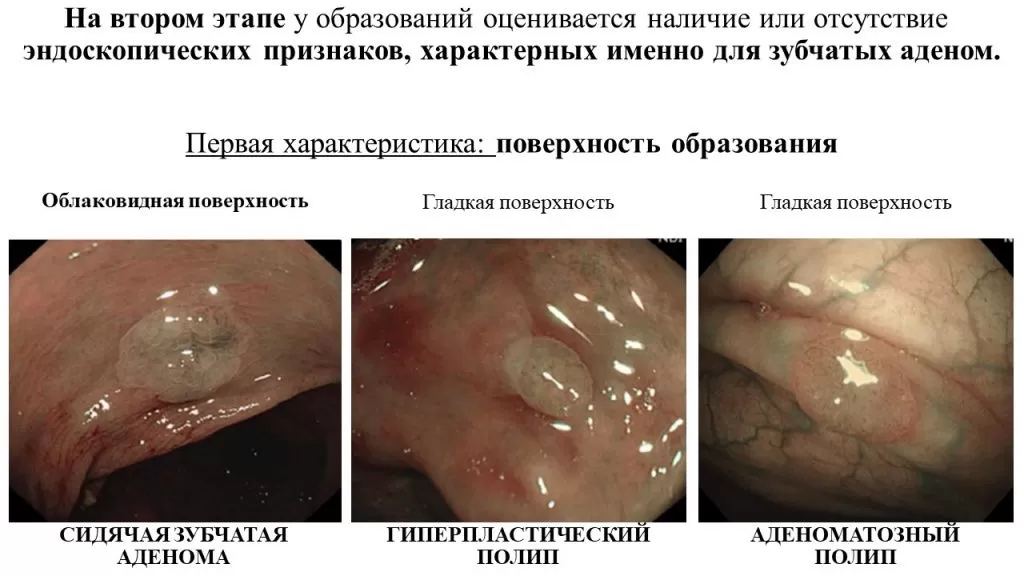
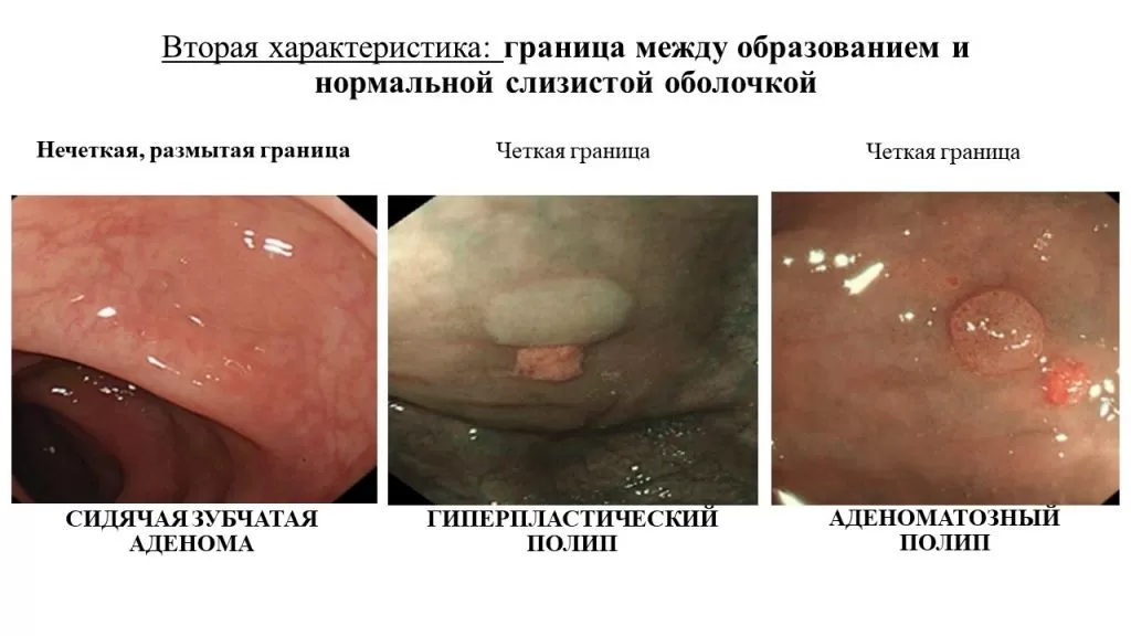
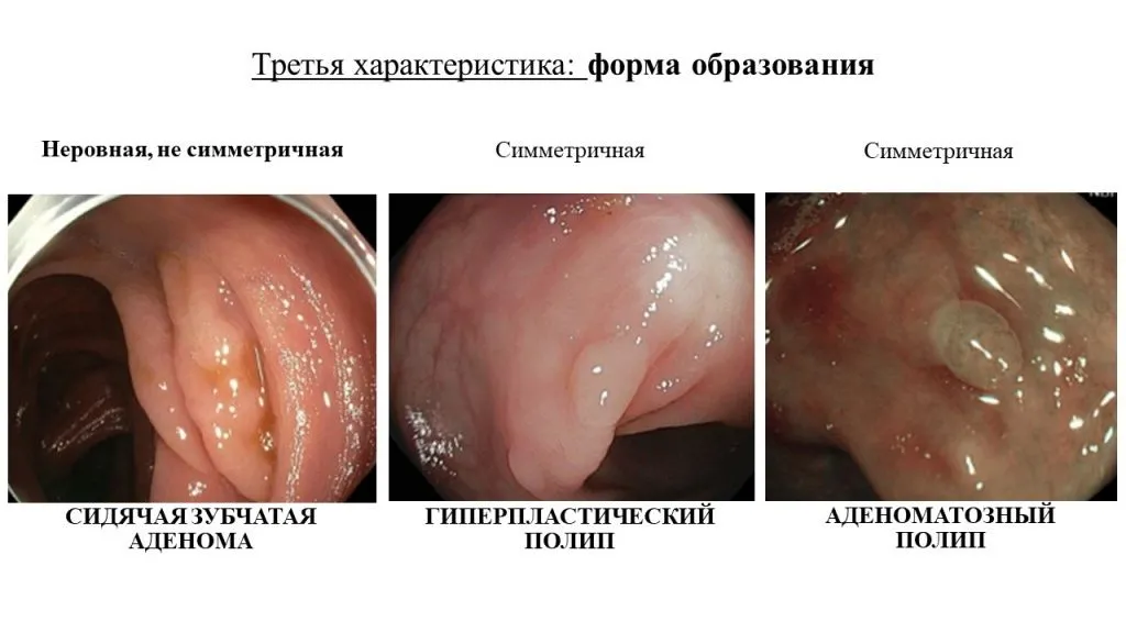
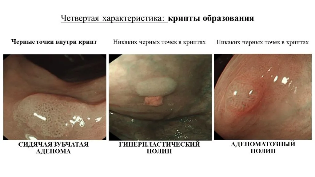
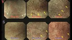
На втором этапе у данных образований оценивается наличие или отсутствие эндоскопических признаков, характерных именно для зубчатых аденом [8].
К ним относятся:
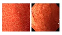
- 1. облаковидная поверхность,
- 2. нечеткая граница,
- 3. неправильная форма,
- 4. «черные точки» внутри крипт (ямки IIo типа по T.Kimura).
Исходя из сочетания эндоскопических признаков делается вывод о предполагаемой морфологической структуре образования.
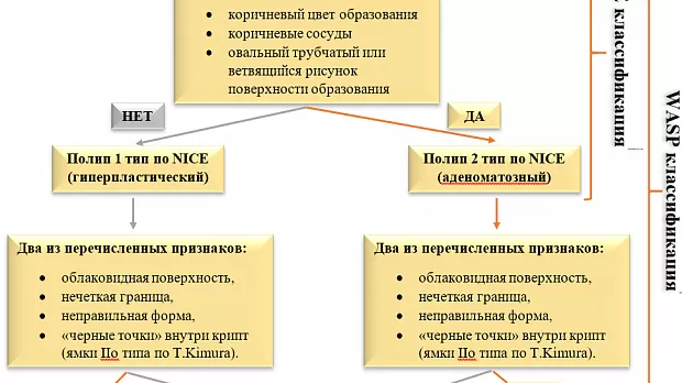
Структура дифференицально-диагностического подхода изображена на схеме:
Надпись: NICE классификация ,Надпись: WASP классификация
|
Один из перечисленных признаков: · коричневый цвет образования · коричневые сосуды · овальный трубчатый или ветвящийся рисунок поверхности образования
| |||
|
|
| ||
|
НЕТ |
ДА | ||
|
Полип 1 тип по NICE (гиперпластический) |
Полип 2 тип по NICE (аденоматозный)
| ||
|
|
| ||
|
Два из перечисленных признаков: · облаковидная поверхность, · нечеткая граница, · неправильная форма, · «черные точки» внутри крипт (ямки IIo типа по T.Kimura).
|
Два из перечисленных признаков: · облаковидная поверхность, · нечеткая граница, · неправильная форма, · «черные точки» внутри крипт (ямки IIo типа по T.Kimura).
| ||
|
|
|
|
|
|
НЕТ |
ДА |
ДА |
НЕТ |
|
|
|
| |
|
Гиперпластический полип |
Зубчатая аденома |
Обычная аденома | |
Для лучшего понимания и запоминания материала предлагаем Вам попрактиковаться в применении данного алгоритма!
По теме:
- ВИЗУАЛЬНАЯ ДИАГНОСТИКА ЗУБЧАТЫХ ОБРАЗОВАНИЙ ТОЛСТОЙ КИШКИ
- Стратегии клинического наблюдения после эндоскопического лечения зубчатых аденом толстой и прямой кишки
- Видео: BASIC (BLI Adenoma Serrated International Classification) классификация колоректальных полипов Video
- Классификация WASP: видео-практикум
- NICE - International Colorectal Endoscopic classification NBI — объединённая эндоскопическая классификация колоректальных неоплазий
Список литературы:
От редакции EndoExpert.ru
- классификации в эндоскопии
- Азбука Эндоскописта. Эндоскопические классификации для описания и оценки патологических изменений толстой кишки при колоноскопии
- Классификация WASP: дифференциальная диагностика гиперпластических полипов, зубчатых аденом и обычных аденом толстой кишки
- Классификация и диагностика гастритов и гастропатий
- Номенклатурная классификация медицинских изделий 3.15 Эндоскопы гастроэнтерологические
- Z-линия пищевода. Классификация ZAP
- Парижская классификация
- Классификации ЭУС критериев хронического панкреатита
Статьи по теме
Рекомендуемые статьи
Эластография при эндосонографии (ЭУС или эндоУЗИ)
Эластография (эластосонография) – метод виртуальной пальпации (технология улучшенной визуализации при ЭУС диагностике), позволяющий дифференцировать злокачественные и доброкачественные поражения лимфоузлов. Основана на принципе, что более мягкие ткани при сжатии легче деформируются, это позволяет объективно оценить консистенцию ткани, показать различия в плотности между нормальными и патологически измененными тканями.
При эндоскопическом исследовании в случае бронхоэктазов в стадии ремиссии выявляется
частично диффузный бронхит I степени воспаления
Активируйте PUSH уведомления в браузер
Отключите PUSH уведомления в браузер
Содержание
Интернет магазин
Популярное
- О нас
- Правовые вопросы
- Политика
обработки персональных
данных EndoExpert.ru - Связаться с нами
- Стать партнером
© 2016-2022 EndoExpert.ru

Вы находитесь в разделе предназначенном только для специалистов (раздел для пациентов по ссылке). Пожалуйста, внимательно прочитайте полные условия использования и подтвердите, что Вы являетесь медицинским или фармацевтическим работником или студентом медицинского образовательного учреждения и подтверждаете своё понимание и согласие с тем, что применение рецептурных препаратов, обращение за той или иной медицинской услугой, равно как и ее выполнение, использование медицинских изделий, выбор метода профилактики, диагностики, лечения, медицинской реабилитации, равно как и их применение, возможны только после предварительной консультации со специалистом. Мы используем файлы cookie, чтобы предложить Вам лучший опыт взаимодействия. Файлы cookie позволяют адаптировать веб-сайты к вашим интересам и предпочтениям.
Я прочитал и настоящим принимаю вышеизложенное, хочу продолжить ознакомление с размещенной на данном сайте информацией для специалистов.





.jpg)
.png)



Комментарии 1